Lower Energy.
Conventional mechanical ventilation generates more power than is needed to induce inspiration and expiration. The net overspill of energy is dissipated in the lungs, which has been shown even when applied for only a few hours to be a source of lung injury, so-called ‘ventilator-induced lung injury’ (VILI).
Recently, clear theoretical evidence was provided for lower energy dissipation in the lungs by FCV® as compared to VCV or PCV. A relatively simple analysis and numerical calculations indicated that energy dissipation is minimized by controlling the ventilation flow to be constant and continuous during both inspiration and expiration, and by ventilating at an I:E ratio close to 1:1. In other words, by using FCV®
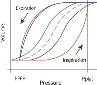
Energy dissipation can be calculated from the hysteresis area of pressure-volume loops obtained during ventilation. PV loops calculated based on routine ventilation protocols showed a 53% reduction in energy dissipation by FCV® as compared to PCV and a 32% reduction as compared to VCV.
Additionally, it was emphasized that accurate measurement of intratracheal pressures is crucial for calculating energy dissipation. Where other VCV and PCV ventilators rely on calculated airway pressures, Evone is the only device that actually measures intratracheal pressures and is thus capable of measuring energy dissipation accurately.
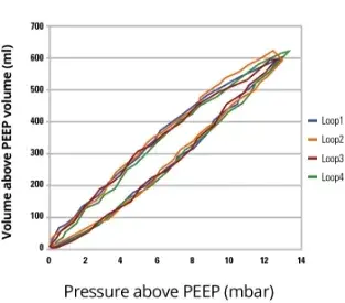
This theory was further validated on a patient. Pressure-volume (PV) loops were recorded in real time, and the energy dissipated in the patient’s lungs was calculated from the hysteresis area of the PV loops. Strikingly, the energy dissipation was just 0.17 J/L, which is even lower than values reported for spontaneous breathing (0.2–0.7 J/L).

Idealized PV loops (the enclosed area of each loop is the dissipated energy) during PCV (red line), VCV (blue line) and FCV® (blue line during inspiration, green line during expiration). The dashed line is the static compliance curve of the lung/chest sys

Real-time measured PV loops of a patient ventilated with FCV® demonstrating minimized hysteresis area of the PV loops (=energy dissipation). (Adopted from Barnes and Enk, TACC 2019 24).


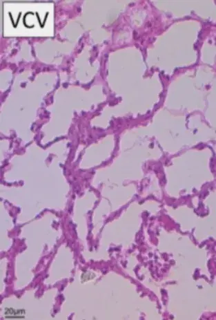
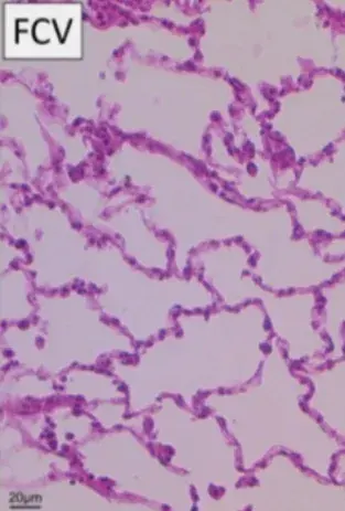
Stained lung tissue samples retrieved after VCV or FCV® ventilation of ARDS pigs, revealing less thickening of alveolar walls in FCV® group, cell infiltration was lower, and surfactant protein A concentration was higher in the FCV® group, indicating the potential for FCV® to attenuate lung injury and to provide lung-protective effects.(Adapted from Schmidt et al. 2020 10).
Benefits of Lower Energy.
The following benefits as compared to Volume Controlled Ventilation (VCV) and Pressure Controlled Ventilation (PCV) may be expected while ventilating patients in FCV® mode:
- FCV® results in smooth tidal movements of the diaphragm and thoracic wall throughout the ventilation cycle.
- FCV® controls expiration and prevents abrupt airway pressure drop.
- Relies on accurately measured intratracheal pressures and inspiratory flows, allowing precise calculation of energy dissipation 23, 24.
- Hysteresis area of pressure-volume loops reflects the energy dissipated 23.
- Constant gas flow in combination with an I:E ratio of 1:1 minimizes energy dissipation down to values reported for spontaneous breathing 23.
- Provides ventilation with reduced mechanical power compared to VCV and PCV. 5, 10, 16, 23, 24.
- Has lung-protective potential 1, 4, 5, 8, 10, 14, 16, 18, 20, 22-24.